

Lab-Grown Organs and Bioengineered Tissue: Advances Toward Transplantable Artificial Organs
Researchers are advancing bioengineered, vascularized tissues to overcome blood supply challenges and permanently change organ transplantation.
In Brief
- Organ shortages leave thousands of patients waiting for lifesaving transplants each year.
- Scientists are developing lab-grown tissues and organs through regenerative medicine and bioengineering.
- One major challenge is creating blood vessel networks that can keep engineered tissues alive.
- Recent breakthroughs in vascularized tissues are bringing researchers closer to transplantable artificial organs.
- These advances could transform transplantation, personalized medicine, and future healthcare.
A patient waiting for a heart, kidney, or liver transplant may spend months or even years hoping a donor organ becomes available. For many patients, that organ never comes. Scientists are now working toward a different solution: growing replacement tissues and organs in the laboratory through regenerative medicine and bioengineering.
Organ transplantation has saved millions of lives, but the global demand for donor organs continues to exceed the available supply. According to Martin et al. (2025), thousands of patients worldwide remain dependent on transplant waiting lists because the amount of donated organs cannot meet the medical demand. This global shortage is reflected in the United States, where tens of thousands of patients continue to wait for lifesaving organ transplants each year, as shown in Figure 1.
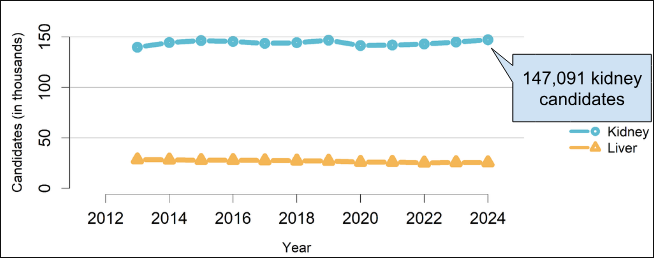
Figure 1. Kidney and liver transplant waiting lists in the United States (2013–2024). The number of patients awaiting kidney and liver transplantation remained consistently high over the past decade, reflecting the persistent demand for donor organs. Adapted from OPTN/SRTR (2024).
Because donor organs remain in critically short supply, scientists are developing new ways to create replacement tissues in the laboratory. One promising solution is regenerative medicine, which has allowed researchers to create increasingly sophisticated laboratory-grown tissues. Where scientists were once limited to growing simple layers of cells, they can now develop organoids, tissue-engineered structures, tissue patches, organ-on-chip systems, and 3D bioprinted tissues that mimic certain features of human organs. Advances in tissue engineering and 3D bioprinting have allowed scientists to precisely arrange cells and biomaterials into organized patterns, making engineered tissues more biologically realistic. These technologies have improved disease modeling, drug testing, tissue repair research, and the development of potential future therapies. However, fully functional transplantable organs have not yet been achieved. One of the greatest challenges is vascularization—the development of blood vessel networks capable of delivering oxygen and nutrients throughout large engineered tissues to keep them alive.
One of the most active areas of research focuses on vascularization, or building blood vessel networks within engineered tissues. Recent advances in this area have significantly improved scientists' ability to keep larger lab-grown tissues alive.
How Scientists Build Living Tissues
The foundation of many regenerative medicine technologies is the use of stem cells. Stem cells are unique because they can develop into specialized cell types found throughout the human body. Depending on the signals they receive, stem cells can become heart cells, liver cells, kidney cells, nerve cells, and many other types of tissue. This flexibility makes them an important tool for researchers attempting to create replacement tissues and study how organs develop.
To build living tissues, scientists use a process known as tissue engineering, as illustrated in Figure 2. Tissue engineering combines biology, engineering, and materials science to create structures that imitate natural tissues. Researchers often begin with stem cells because they can develop into the specialized cells needed for different tissues. These cells are combined with biomaterials that provide structural support and engineered scaffolds that organize them into specific shapes. The cells then multiply and mature, gradually forming living tissue within the scaffold. Once the engineered tissue becomes larger and more complex, however, cells located farther from the surface can no longer receive enough oxygen and nutrients through diffusion alone.
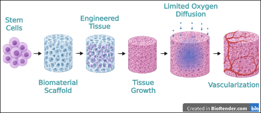
Figure 2. Tissue engineering workflow illustrating the progression from stem cells to engineered tissue. As engineered tissues increase in size, oxygen diffusion becomes limited, creating the need for vascularization. Created with BioRender.com.
This limitation creates one of the greatest obstacles to developing larger artificial tissues and organs: vascularization, the formation of blood vessels that deliver oxygen and nutrients throughout the tissue. All living tissues require a constant supply of oxygen and nutrients to survive. In the human body, blood vessels perform this function by transporting oxygen and nutrients to cells while removing waste products. Without a functional blood vessel network, cells located deep within engineered tissues cannot receive the resources they need to survive.
As engineered tissues increase in size, this problem becomes even more significant. Small tissue structures may survive through simple diffusion of oxygen and nutrients, but larger tissues require their own circulation systems. Creating stable blood vessel networks capable of supporting complex tissues remains one of the greatest challenges in regenerative medicine and a critical step toward developing fully transplantable artificial organs.
Breakthrough Story: Solving the Vascularization Challenge
For years, scientists could grow human cells and create small pieces of tissue in the laboratory. The real challenge began when they tried to make those tissues larger. Cells deep inside engineered tissues often died because oxygen and nutrients could not reach them.
Researchers soon realized that building an artificial organ would require much more than growing the right cells. Every organ in the human body depends on an extensive network of blood vessels that continuously delivers oxygen and removes waste. Without a similar system, lab-grown tissues could never survive long enough to function as replacement organs.
Early tissue-engineering efforts focused on creating simple tissue structures and developing scaffolds that could support growing cells. These approaches helped researchers organize cells into more realistic arrangements, but they still could not reproduce the circulation systems found in natural organs.
To address this problem, scientists began designing artificial vascular channels—small pathways that mimic blood vessels. Advances in biomaterials allowed these channels to be incorporated into engineered tissues, improving the movement of nutrients and oxygen throughout the structure.
A major turning point came with the development of 3D bioprinting. Instead of allowing cells to grow randomly, researchers could precisely place cells and biomaterials into specific patterns. Scientists began printing tissues with embedded vascular channels, bringing engineered tissues one step closer to functioning like natural organs.
One example is the Sacrificial Writing Into Functional Tissue (SWIFT) biomanufacturing process, shown in Figure 3, which uses a temporary "sacrificial" ink to create hollow vascular channels within densely packed stem cell–derived tissues. After the ink is removed, these channels can be perfused with oxygen and nutrients, helping engineered tissues survive and function more like natural organs.
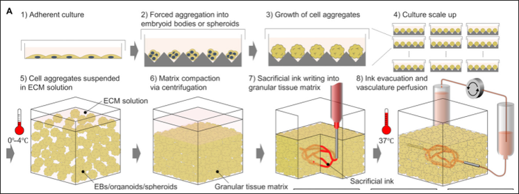
Figure 3. SWIFT biomanufacturing process for vascularized engineered tissue. Stem cell–derived organoids are compacted into a living tissue matrix, where sacrificial bioprinting creates hollow vascular channels for tissue perfusion. This approach helps overcome one of the major challenges in engineering transplantable tissues. Adapted from Skylar-Scott et al. (2019).
Recent advances have accelerated progress even further. Researchers have developed vascularized organoids-on-chip (Figure 4) that combine miniature organ models with functional circulation systems, allowing tissues to behave more like those found in the human body. These systems use several vascularization strategies, including endothelial cell coculture, gene overexpression, organoid fusion, and microfluidic perfusion to support blood vessel formation. Other teams have also reported implantable vascularized tissue constructs designed to promote blood vessel growth after transplantation. These advances provide researchers with new ways to study how engineered tissues survive, grow, and interact with living environments.
One of the most ambitious projects currently underway is a Stanford-led effort to bioprint a human heart and eventually implant it into a pig. Researchers are combining stem-cell biology, tissue engineering, and advanced bioprinting technologies to create heart tissue with the vascular networks needed to keep it alive. Although fully transplantable artificial hearts remain a long-term goal, projects like this demonstrate how rapidly the field is advancing.
Taken together, these advances illustrate a clear progression in regenerative medicine. Researchers first addressed the challenge of keeping engineered tissues alive by developing artificial vascular channels and later refined these approaches through 3D bioprinting and vascularized organoids-on-chip. Today, ambitious projects such as Stanford's effort to bioprint a human heart demonstrate how these innovations are bringing scientists closer to creating transplantable artificial organs.
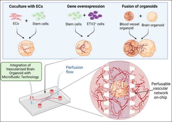
Figure 4. Strategies for vascularizing engineered organoids. Endothelial cell coculture, gene overexpression, organoid fusion, and microfluidic organoid-on-chip technology promote blood vessel formation and improve oxygen and nutrient delivery within engineered tissues. Adapted from Tan et al. (2023).
Remaining Challenges
Although advances in vascularized tissue engineering have brought scientists closer to creating artificial organs, several major challenges must still be overcome before these technologies can become routine medical treatments. Successfully growing living tissue in the laboratory is only the first step; researchers must also ensure that engineered organs can be produced at a practical scale, remain affordable, function safely in patients, and meet strict regulatory standards.
One of the greatest challenges is scalability. Creating small engineered tissues is much easier than producing full-sized organs with the same level of complexity found in the human body. Natural organs contain billions of cells organized into highly structured systems, and reproducing this organization on a large scale remains a major difficulty.
Even if scientists can overcome this challenge, cost remains another significant barrier. Many regenerative medicine technologies require advanced equipment, specialized materials, and highly trained researchers. The resources needed to create and test engineered tissues can be expensive, meaning that reducing costs will be necessary before these treatments can become accessible to more patients.
Beyond manufacturing and cost, researchers must also demonstrate that engineered organs are safe for long-term use. Before these tissues can be implanted into patients, scientists must prove that they can function properly over extended periods and interact safely with the human body. Continued research is needed to understand how engineered tissues perform after transplantation and how the body responds to them.
Finally, even after these scientific and medical challenges are addressed, engineered organs must undergo extensive regulatory review before they can be used in clinical practice. Because these technologies combine biology, engineering, and medicine, they must be thoroughly evaluated to demonstrate their safety and effectiveness before receiving approval for clinical applications.
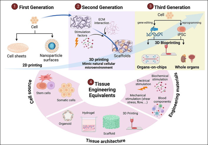
Figure 5. Evolution of tissue engineering technologies from early cell-based approaches to modern regenerative medicine strategies, including organoids, 3D bioprinting, organ-on-a-chip systems, and whole-organ engineering. Adapted from Hoang et al. (2025).
Why This Matters: Future Implications and Career Connections
The development of lab-grown tissues and artificial organs has the potential to transform modern medicine. One of the most significant benefits could be a reduced dependence on donor organs. If scientists can successfully create transplantable organs in the laboratory, patients may no longer need to spend years on transplant waiting lists or rely solely on donor availability. This could improve access to lifesaving treatments and help address the persistent shortage of donor organs.
These technologies may also advance personalized medicine. Researchers are exploring ways to create engineered tissues using a patient's own cells, which could reduce the risk of immune rejection after transplantation. Personalized tissues may allow treatments to be tailored to individual patients, improving both safety and effectiveness.
Beyond transplantation, engineered tissues already play an important role in biomedical research. Organoids and other laboratory-grown tissue models allow scientists to study diseases and test potential treatments using systems that more closely resemble human biology. As these technologies continue to improve, they may help researchers develop safer and more effective therapies while reducing reliance on traditional testing methods.
The impact of regenerative medicine extends beyond transplant surgery. As scientists improve their ability to engineer functional tissues, future treatments may be able to repair or replace damaged organs instead of relying solely on donor transplantation. These advances could provide new treatment options for patients with severe injuries, congenital defects, and diseases that currently have few long-term solutions, fundamentally changing how many conditions are treated and improving long-term patient outcomes.
Researchers continue to refine the technologies discussed throughout this article. One major area of focus is improving vascularization so engineered tissues can develop stable blood vessel networks capable of supplying oxygen and nutrients after transplantation. Scientists are also advancing 3D bioprinting techniques to create increasingly complex tissues with more accurate biological structures. Continued progress in these areas will be essential for producing fully transplantable artificial organs that function like their natural counterparts.
The field of regenerative medicine also creates opportunities for a wide range of careers. Professionals in biomedical engineering, biotechnology, tissue engineering, clinical research, and regenerative medicine all contribute to the development of new therapies and technologies. Biomedical engineers design scaffolds and bioprinting systems, tissue engineers develop laboratory-grown tissues, clinical researchers evaluate the safety and effectiveness of new treatments, and biotechnology professionals help translate laboratory discoveries into therapies that can be manufactured and used in healthcare. Collaboration among these fields is essential for bringing engineered organs from the laboratory to patients.
If you've ever been interested in biology, engineering, medicine, or even computer design, regenerative medicine brings all of these fields together. Teams developing artificial organs often include biomedical engineers who design scaffolds and bioprinting systems, surgeons who determine how engineered organs can be safely transplanted into patients, stem-cell researchers who develop the living cells used to create tissues, software specialists who create imaging and bioprinting programs that guide tissue construction, and materials scientists who develop biomaterials that support cell growth and tissue formation. Together, these specialists combine their expertise to move regenerative medicine from laboratory research toward real-world medical treatments. The next generation of breakthroughs may come from students who choose to pursue careers in these fields.
Ultimately, the long-term implications of this research extend beyond individual technologies. As scientists improve their ability to create functional tissues and organs, medicine may gradually shift from relying primarily on organ donation toward manufacturing personalized replacement tissues. Although significant challenges remain, current advances suggest that this possibility is becoming increasingly realistic.
Conclusion
A decade ago, scientists could grow only small, simple tissues in the laboratory, and creating fully functional organs seemed like science fiction. As researchers developed artificial vascular networks, advanced 3D bioprinting techniques, and increasingly sophisticated organ-on-chip systems, each breakthrough brought them one step closer to overcoming the challenges that once limited the field. Today, the goal of creating transplantable artificial organs no longer feels impossible—it represents a rapidly advancing area of scientific research.
Significant challenges remain, including scalability, cost, safety, and regulatory approval. However, the progress made in vascularized tissue engineering demonstrates how quickly regenerative medicine is advancing. What once seemed impossible is gradually becoming a realistic scientific goal.
For students interested in medicine, biotechnology, engineering, or research, this field offers a glimpse into how scientific innovation can directly improve human lives. As scientists continue developing ways to create functional replacement tissues and organs, the future of healthcare may look very different from the one we know today.
References
- Alkazemi, H., Mitchell, G. M., Lokmic-Tomkins, Z., Heath, D. E., & O'Connor, A. J. (2024). Hierarchically vascularized and implantable tissue constructs created through angiogenesis from tissue-engineered vascular grafts. bioRxiv. https://www.biorxiv.org/content/10.1101/2024.04.29.591796v1
- Hoang, V. T., Nguyen, Q. T., Phan, T. T. K., Pham, T. H., Dinh, N. T. H., Anh, L. P. H., Dao, L. T. M., Bui, V. D., Dao, H.-N., Le, D. S., Ngo, A. T. L., Le, Q.-D., & Nguyen Thanh, L. (2025). Tissue engineering and regenerative medicine: Perspectives and challenges. MedComm, 6(5), e70192. https://doi.org/10.1002/mco2.70192
- Lewis Lab. (n.d.). Three-dimensional bioprinting of thick vascularized tissues. Harvard John A. Paulson School of Engineering and Applied Sciences. https://lewisgroup.seas.harvard.edu/publications/three-dimensional-bioprinting-thick-vascularized-tissues
- Martin, F., Carmona, M., Mahillo, B., Alvarez, M., Luengo, A., Chatzixiros, E., López-Fraga, M., Domínguez-Gil, B., & Tullius, S. G. (2025). Global organ donation and transplantation activities, 2024. Transplant International. https://pmc.ncbi.nlm.nih.gov/articles/PMC12908642/
- OPTN/SRTR. (2024). 2024 annual data report: Overview of U.S. solid organ transplantation. https://srtr.hrsa.gov/adr/2024/Overview/
- Skylar-Scott, M. A., et al. (2023, September 12). Moonshot effort aims to bioprint a human heart and implant it in a pig. Stanford Report. https://news.stanford.edu/stories/2023/09/moonshot-effort-aims-bioprint-human-heart-implant-pig
- Skylar-Scott, M. A., Uzel, S. G. M., Nam, L. L., Ahrens, J. H., Truby, R. L., Damaraju, S., & Lewis, J. A. (2019). Biomanufacturing of organ-specific tissues with high cellular density and embedded vascular channels. Science Advances, 5(9), eaaw2459. https://www.science.org/doi/10.1126/sciadv.aaw2459
- Tan, S. Y., Feng, X., Cheng, L. K. W., & Wu, A. R. (2023). Vascularized human brain organoid on-chip. Lab on a Chip, 23(12), 2693–2709. https://doi.org/10.1039/d2lc01109c
- U.S. Department of Health & Human Services. (n.d.). Organ donation statistics. OrganDonor.gov. https://www.organdonor.gov/learn/organ-donation-statistics/detailed-description
- Zhao, Y., Landau, S., Okhovatian, S., et al. (2024). Integrating organoids and organ-on-a-chip devices. Nature Reviews Bioengineering, 2, 588–608. https://doi.org/10.1038/s44222-024-00207-z